41 microscope labels and definitions
Labeling the Parts of the Microscope Labeling the Parts of the Microscope This activity has been designed for use in homes and schools. Each microscope layout (both blank and the version with answers) are available as PDF downloads. You can view a more in-depth review of each part of the microscope here. Download the Label the Parts of the Microscope PDF printable version here. › books › NBK310485Laboratory procedures for diagnosis of anthrax, and isolation ... 1. Anthrax and the microbiology laboratory; operational safety. With some country-to-country variation in safety level definitions and requirements, recommendations for the manipulation of the causative agent of anthrax, Bacillus anthracis, generally are that BSL (biosafety level) 2 practices, containment equipment and facilities are appropriate for diagnostic tests, but BSL3 standards should ...
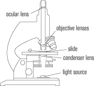
Parts of the Microscope with Labeling (also Free Printouts) Parts of the Microscope with Labeling (also Free Printouts) A microscope is one of the invaluable tools in the laboratory setting. It is used to observe things that cannot be seen by the naked eye. Table of Contents 1. Eyepiece 2. Body tube/Head 3. Turret/Nose piece 4. Objective lenses 5. Knobs (fine and coarse) 6. Stage and stage clips 7. Aperture
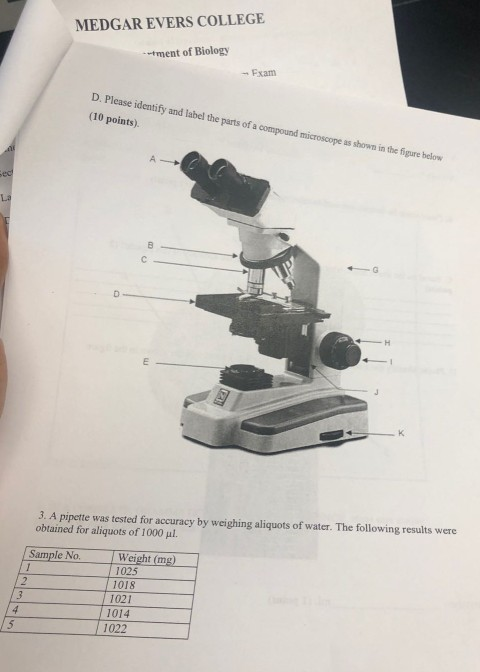
Microscope labels and definitions
Parts of the Microscope Label and Definition Diagram - Quizlet Parts of the Microscope Label and Definition STUDY Learn Flashcards Write Spell Test PLAY Match Gravity Created by jlabel Terms in this set (14) Body Tube Keeps correct distance between eyepiece and lens Revolving Nosepiece Holds high and low power objectives, can be rotated to adjust magnification Low Power Objective › obp › uiISO 18113-1:2009(en), In vitro diagnostic medical devices ... However, definitions provided in national and regional regulations shall take precedence. Furthermore, while the terms and definitions in International Standards are preferred, the terms and definitions used in the information supplied by an IVD manufacturer shall be subject to the requirements of 4.6.2. en.wikipedia.org › wiki › Optical_resolutionOptical resolution - Wikipedia In a properly configured microscope, + =. The above estimates of resolution are specific to the case in which two identical very small samples that radiate incoherently in all directions. Other considerations must be taken into account if the sources radiate at different levels of intensity, are coherent, large, or radiate in non-uniform patterns.
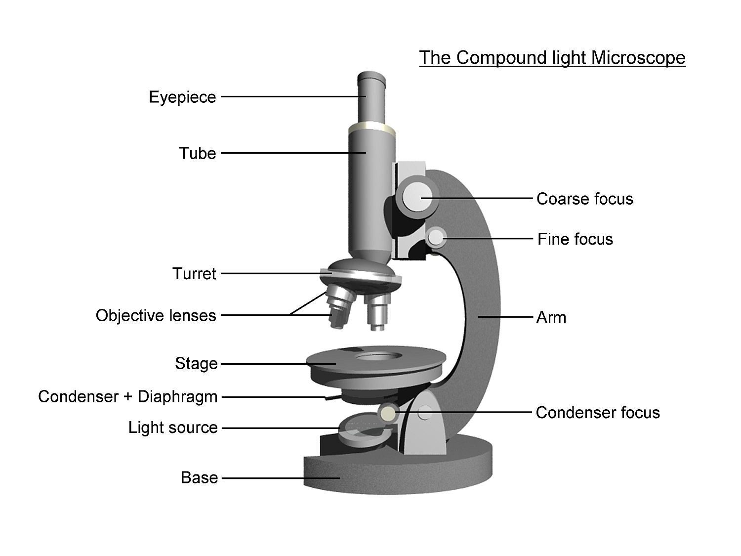
Microscope labels and definitions. Microscope Types (with labeled diagrams) and Functions A compound microscope: Is used to view samples that are not visible to the naked eye. Uses two types of lenses - Objective and ocular lenses. Has a higher level of magnification - Typically up to 2000x. Is used in hospitals and forensic labs by scientists, biologists and researchers to study micro organisms. Compound microscope labeled diagram. PDF Definitions of the Parts of the Microscope - UAlberta microscope moves the stage up and down to bring the specimen into focus. The gearing mechanism of the adjustment produces a large vertical movement of the stage with only a partial revolution of the knob. Because of this, the coarse adjustment should only be used with low power (4X and 10X objectives) and never with the high power lenses (40X and Microscope: Definition, Types, Uses, Parts & Examples | Toppr 4. Fluorescence Microscope. These scopes use ultraviolet light to illuminate specimens that fluoresce. Besides, mostly, a fluorescent antibody or dye is added on the viewed specimen. 5. Contrast/Phase Microscope. This scope uses a special condenser that allows the examination of structures inside the cells. Also, they use a compound light. Microscope Parts and Functions First, the purpose of a microscope is to magnify a small object or to magnify the fine details of a larger object in order to examine minute specimens that cannot be seen by the naked eye. Here are the important compound microscope parts... Eyepiece: The lens the viewer looks through to see the specimen.
Label Microscope Diagram - EnchantedLearning.com low-power objective - a small lens with low magnifying power. mirror (or light source) - this directs light upwards onto the slide. revolving nosepiece - the rotating device that holds the objectives (lenses). stage - the platform on which a slide is placed. stage clips - metal clips that hold a slide securely onto the stage. Advertisement. Compound Microscope Parts, Diagram Definition, Application, Working ... Definition of a Compound Microscope and uses. parts of a compound microscope and application. compound microscope labeled diagram. MN . Generic selectors. ... For future reference, adhesive labels are stuck to the base and sides of the microscope. (iii) Use. After calibrating the eyepiece scales for all objective lenses, the microscope can be ... Parts of Stereo Microscope (Dissecting microscope) - labeled diagram ... Stereo microscopes (also called Dissecting microscope) are branched out from other light microscopes for the application of viewing "3D" objects. These include substantial specimens, such as insects, feathers, leaves, rocks, sand grains, gems, coins, and stamps, etc. Functionally, a stereo microscope is like a powerful magnifying glass. PDF Parts of a Microscope Printables - Homeschool Creations Label the parts of the microscope. You can use the word bank below to fill in the blanks or cut and paste the words at the bottom. Microscope Created by Jolanthe @ HomeschoolCreations.net. Parts of a eyepiece arm stageclips nosepiece focusing knobs illuminator stage objective lenses
& Barcode Equipment | Adazon Adazon is the leading provider of labeling and barcode equipment online. We offer custom labels, printers, software, point of sale kits and more. Order today! Microscope, Microscope Parts, Labeled Diagram, and Functions Microscopes magnify or enlarge small objects such as cells, microbes, bacteria, viruses, microorganisms etc. at a viewable scale for examination and analysis. Microscopes consist of one or more magnification lenses to enlarge the image of the microscopic objects placed in the focal plane. thegradient.pub › interpretability-in-ml-a-broadInterpretability in Machine Learning: An Overview - The Gradient Nov 21, 2020 · Murdoch, W. James, et al. "Definitions, methods, and applications in interpretable machine learning." Proceedings of the National Academy of Sciences 116.44 (2019): 22071-22080. Roscher, Ribana, et al. "Explainable machine learning for scientific insights and discoveries." IEEE Access 8 (2020): 42200-42216. Microscope Glossary A microscope is typically composed of a head or body and a base. The base is the support mechanism. Binocular Microscope A microscope with a head that has two eyepiece lens. Nowadays, binocular is typically used to refer to compound or high power microscopes where the two eyepieces view through a single objective lens.
32 Label Of Compound Microscope - Label Design Ideas 2020
Compound Microscope Parts, Functions, and Labeled Diagram Compound Microscope Definitions for Labels. Eyepiece (ocular lens) with or without Pointer: The part that is looked through at the top of the compound microscope. Eyepieces typically have a magnification between 5x & 30x. Monocular or Binocular Head: Structural support that holds & connects the eyepieces to the objective lenses.

Animal Cell Quizlet With Pictures : 7th Grade Microscope Cell Osmosis Diffusion Flashcards ...
Microscope Glossary of Terms: Microscope A-Z - Microscope and ... Body - The upper part of the microscope including the stage and is often referred to alongside the eyepiece. Body Tube Length - This refers to the distance between the objective and the very top of the body tube. This can be important as objective lenses are compatible with certain body tube lengths and a mismatch can cause spherical aberrations.

Label the Microscope Part
Microscope Glossary of Terms - OpticsPlanet 9. Base. The part of the microscope that comes in contact with the table or other surface used to support it. 10. Bertrand Lens. This is a small lens used in the tube of a polarized light microscope and is used to study interference patterns for the sake of identification and analysis. 11.
30 Label The Indicated Parts Of The Microscope - Label Ideas 2020
Glossary of Widely Used Microscopy Terms and Imaging Techniques - ZEISS Geometric deviation of an image formed by an imaging grating from the ideal point image. Absorbance A.- The logarithm to the base 10 of the reciprocal of the transmittance, (T). A = log10 (1/T) = - log10 T Absorption Band A region of the absorption spectrum in which the absorbance passes through a maximum. ACE Automatic Component Extraction
Microscope Quiz
Microscope Terms | Microscope World Resources Articulated Arm: A type of microscope stand that holds a microscope body. The stand clamps to a table or has a large base and has a variety of motion in three dimensions. The microscope body is held onto the articulated arm stand with a focusing holder. Articulated arm microscope stands are often used in industrial and manufacturing settings.
microscope - DriverLayer Search Engine
Microscope Parts & Functions - AmScope Invented by a Dutch spectacle maker in the late 16th century, compound light microscopes use two sets of lenses to magnify images for study and observation. The first set of lenses are the oculars, or eyepieces, that the viewer looks into; the second set of lenses are the objectives, which are closest to the specimen.

Mshot MC20-C Microscopes Camera 140 mega pixel | eBay
Compound Microscope Parts - Labeled Diagram and their Functions - Rs ... A microscope is an instrument used to see objects that are too small to be seen by the naked eye. We have an article covering the history, types, and evolution of all kinds of microscopes. If you are interested in this topic, please click the link above. [In this figure] The name "microscope" came from two words - "micro" and "scope".
The compound microscope uses ~ Facebook-emo
› books › NBK21116Mapping Genomes - Genomes - NCBI Bookshelf These labels combine high sensitivity with high resolution and are ideal for in situ hybridization. Fluorolabels with different colored emissions have been designed, making it possible to hybridize a number of different probes to a single chromosome and distinguish their individual hybridization signals, thus enabling the relative positions of ...

Microscope - diagram Tom Butler | Technical Drawing and Illustration Projects | Pinterest ...
Microscope Magnification: Explained - Microscope Clarity To calculate the magnification on a microscope multiply the magnification power of the eyepiece you are using by the objective currently in position. Magnification = Eyepiece Magnification X Objective Magnification. Microscopes magnify or enlarge the image under inspection and enables the human eye to see things we would never be able to see.

31 Label Of A Microscope - Label Design Ideas 2020
Parts of a microscope with functions and labeled diagram Microscope Definition Microscopes are instruments that are used in science laboratories to visualize very minute objects such as cells, and microorganisms, giving a contrasting image that is magnified. Microscopes are made up of lenses for magnification, each with its own magnification powers.
Label a microscope - Teaching resources
en.wikipedia.org › wiki › DenimDenim - Wikipedia Denim under a microscope. Selvedge identifier visible in white at the interior of a pair of jeans Most denim made today is made on a shuttleless loom [12] that produces bolts of fabric 60 inches (1,500 mm) or wider, but some denim is still woven on the traditional shuttle loom , which typically produces a bolt 30 inches (760 mm) wide.

Science labs - Akers 6th Grade Team
Microscope- Definition, Parts, Functions, Types, Diagram, Uses It is a type of fluorescence microscope that is used to produce 2-D or 3-D images of relatively thick specimens. In this type, the excitation light is focused on a specific spot of sample lying on the focal plane. The focus spot is optically manipulated to scan the entire sample and generate a 3-D image.

Used KEYENCE VE-9800 series 3D high-definition electronic scanning microscope | eBay
Microscope Parts - definitions Flashcards | Quizlet Moves Body Tube up and down for focusing Arm supports body tube Fine Adjustment Sharpens image by moving body tube slightly Nosepiece Holds high and low power objectives, can be rotated to adjust magnification High Power Objective Provides higher magnification, usually about 40x Low Power Objective Provides lower magnification, usually about 10x
34 Label Of A Microscope - Labels For You
Types of Microscopes: Definition, Working Principle, Diagram ... There are also microscope types that find application in metallurgy and studying three-dimensional samples. In this article, there are 5 such microscope types that are discussed along with their diagram, working principle and applications. These five types of microscopes are: Simple microscope. Compound microscope.

Microscope and Its Parts ~ Marta's Blog
Microscope Glossary of Terms | Celestron Microscope - an optical instrument used for viewing objects, typically under levels of magnification several hundred times the objects' actual size. There are three basic parts: head, body, arm. Compound microscope - a microscope that combines the power of lenses and light to enlarge the subject being viewed.

Label And Color The Parts Of Both Microscopes - Best Label Ideas 2019
Compound Microscope- Definition, Labeled Diagram, Principle, Parts, Uses Therefore, a microscope can be understood as an instrument to observe tiny elements. The optical microscope often referred to as the light microscope, is a type of microscope that uses visible light and a system of lenses to magnify images of small subjects. There are two basic types of optical microscopes: Simple microscopes Compound microscopes

Give the label each part of the microscope: - Brainly.ph
Understanding Microscopes and Objectives | Edmund Optics Understanding Microscopes and Objectives. A microscope is an optical device used to image an object onto the human eye or a video device. The earliest microscopes, consisting of two elements, simply produced a larger image of an object under inspection than what the human eye could observe. The design has evolved over the microscope's history ...
Post a Comment for "41 microscope labels and definitions"