45 chlamydomonas diagram with labels
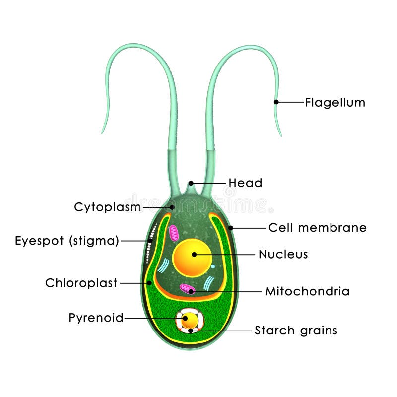
Chlamydomonas reinhardtii - an overview | ScienceDirect Topics Chlamydomonas reinhardtii cells are oval shaped, c. 10 μm in length and 3 μm in width, with two flagellae at their anterior end (Figure 1). The cells contain a single chloroplast occupying 40% of the cell volume and several mitochondria. ... Diagram labeling densities in the averaged image. (B) Image average from thin sections of pf14 ... Navy Removal Scout 800 Pink Pill Assasin Expo Van Travel ... 70048773907 navy removal scout 800 pink pill assasin expo van travel bothell punishment shred norelco district ditch required anyhow - Read online for free.
Biology Diagram Of Chlamydomonas - Which One Of The Following Is A ... Draw A Labeled Diagram Of Chlamydomonas Snapsolve from wb-qb-sg-oss.bytededu.com I tell you about how can we draw labelled diagram of chlamydomonas in . Science, 8th standard text book, ktbs. The thallus is represented by a single cell. Draw chlamydomonas step by step drawing easy to draw#sudhakararts#biological sciences#drawing.

Chlamydomonas diagram with labels
How to make label Diagram of chlamydomonas - YouTube watch: "How to make thumbnail our you tube videos Hindi /urdu haris by #Top2utv" ... Paramecium: Classification, Structure, Diagram, Reproduction by ... - BYJUS Asexual Reproduction in paramecium is by binary fission. The mature cell divides into two cells and each grows rapidly and develops into a new organism. Under favourable conditions, Paramecium multiplies rapidly up to three times a day. Binary fission divides a cell transversely and followed by mitotic division in the micronucleus. Chlamydomonas: Position, Occurrence and Structure (With Diagrams) Chlamydomonas is unicellular, motile green algae. The thallus is represented by a single cell. It is about 20 p,-30|i in length and 20 µ in diameter. The shape of thallus can be oval, spherical, oblong, ellipsoidal or pyriform. The pyriform or pear shaped thalli are common, they have narrow anterior end and a broad posterior end (Fig. 1).
Chlamydomonas diagram with labels. Genetic map of the Chlamydomonas reinhardtii plastid genome ... Download scientific diagram | Genetic map of the Chlamydomonas reinhardtii plastid genome. Protein-coding regions are yellow and their exons are labeled with an "E" followed by a number denoting... Diagram Of Chlamydomonas With Label - Blogger Draw a labelled diagram of chlamydomonas. It is oblong or pyriform in shape. Biological drawings of protista, structure of chlamydomonas,. The anterior end has two tinsel shaped . Shipping a package with ups is easy, as you can print labels for boxes, paste them and even schedule a pickup. Single-cell mass spectrometry - ScienceDirect 11.05.2022 · The labels are used to 'barcode' each sample with stable 13 C and 15 N isotopes arranged at different positions in the linker and reporter groups such that the reporter group provides a readout of the barcode upon collisional activation (MS/MS). (C) For a specific peptide, the MS/MS spectrum can be used to identify the peptide, and the abundances of the reporter … Chlamydomonas - Wikipedia Drawings of Chlamydomonas caudata Wille. [1] Cross section of a Chlamydomonas reinhardtii cell Light micrograph of Chlamydomonas with two flagella just visible at bottom left Chlamydomonas globosa, again with two flagella just visible at bottom left
Substancial | PDF | United Kingdom | Spain - Scribd substancial - Free ebook download as Text File (.txt), PDF File (.pdf) or read book online for free. contains some random words for machine learning natural language processing Solved: Chapter 21 Problem 24TY Solution - Chegg ISBN-13: 9780077388508 ISBN: 007738850X Authors: Sylvia S Mader Rent | Buy. This is an alternate ISBN. View the primary ISBN for: Biology 10th Edition Textbook Solutions. Asymmetric properties of the Chlamydomonas reinhardtii cytoskeleton ... The C. reinhardtii eyespot. (a) A diagram illustrating asymmetric localization of the eyespot relative to the cytoskeleton. Two flagella and four microtubule rootlets extend from a pair of basal bodies at the anterior end of the cell; both the mother basal body (small black oval) and the daughter basal body (small gray oval) are associated with a four-membered rootlet (M4 or D4) and a two ... Finances in Germany - Expat Guide to Germany | Expatica Understanding your money management options as an expat living in Germany can be tricky. From opening a bank account to insuring your family’s home and belongings, it’s important you know which options are right for you.
Root microbiota drive direct integration of phosphate stress and ... 15.03.2017 · In Arabidopsis thaliana, a genetic network that controls the phosphate stress response also influences the structure of the root microbiome community, even under non-stress phosphate conditions. Describe the structure of chlamydomonas with neat labelled diagram ... answeredOct 30, 2020by Naaji(56.8kpoints) selectedOct 30, 2020by Jaini Best answer 1. Chlamydomonas is a simple, unicellular, motile fresh water algae. They are oval, spherical or pyriform in shape. 2. The cell is surrounded by a thin and firm cell wall made of cellulose. 3. The cytoplasm is seen in between the cell membrane and the chloroplast. 4. Substancial | PDF | United Kingdom | Spain - Scribd substancial - Free ebook download as Text File (.txt), PDF File (.pdf) or read book online for free. contains some random words for machine learning natural language processing Spirogyra Labelled Diagram Spirogyra (common names include water silk, mermaid's tresses, and blanket weed) is a genus of filamentous charophyte green algae of the order Zygnematales, named for the helical or spiral arrangement of the chloroplasts that is characteristic of the genus. Draw a labelled diagram of Spirogyra. 51 Differentiate between flying lizard and bird.

Chlamydomonas green algae — Stock Photo #73308961
Chlamydomonas as a Model Organism - Rice University Chlamydomonas as a Model Organism. Chlamydomonas, a genus of unicellular photosynthetic flagellates, is an important model for studies of such fundamental processes as photosynthesis, motility, responses to stimuli such as light, and cell-cell recognition.C. reinhardi, the most commonly studied species of Chlamydomonas, has a relatively simple genome, which has been sequenced.
Chlamydomonas stock illustration. Illustration of algae - 48747331
Diagram of Chlamydomonas angulosa... - Getty Images UNSPECIFIED - CIRCA 2003: Diagram of Chlamydomonas angulosa, Flagellated Protozoan. Drawing. (Photo by DeAgostini/Getty Images)

Phylogenetic analysis supports two evolutionarily conserved tapetal... | Download Scientific Diagram
Life Cycle of Chlamydomonas (With Diagram) - Biology Discussion Each daughter cell develops cell wall, flagella and transforms into zoospore (Fig. 6). The zoospores are liberated from the parent cell or zoosporangium by gelatinization or rupture of the cell wall. The zoospores are identical to the parent cell in structure but smaller in size. The zoospores simply enlarge to become mature Chlamydomonas.

34 Diagram Of Spirogyra With Label - Labels Database 2020
Single-cell mass spectrometry - ScienceDirect May 11, 2022 · The labels are designed to ensure that (i) the total mass of each TMT label (reporter and linker groups) is identical, and (ii) the reporter groups have 18 different masses. Thus, a given peptide ion that is tagged with different labels will have identical masses for m / z selection and ion fragmentation, resulting in abundant sequence ions for ...

DRAW IT NEAT : How to draw Chlamydomonas
Eye Diagram With Labels and detailed description - BYJUS A brief description of the eye along with a well-labelled diagram is given below for reference. Well-Labelled Diagram of Eye The anterior chamber of the eye is the space between the cornea and the iris and is filled with a lubricating fluid, aqueous humour. The vascular layer of the eye, known as the choroid contains the connective tissue.

Vacuole Is Like A Refrigerator Because Everything Forms A...
Actin and Actin-Binding Proteins - PMC The labels are single-letter abbreviations for selected amino acids. ( D ) Cartoon of the actin filament showing the position of the pointed and barbed ends. ( A , B , Reprinted, with permission, from Pollard and Earnshaw 2007 ; C , reprinted, with permission from Macmillan Publishers Ltd., from Fujii et al. 2010 ; D , adapted, with permission, from Pollard and Earnshaw 2007 .)

Algae
Draw a neat labelled diagram. Chlamydomonas - Shaalaa.com Draw a neat labelled diagram. Chlamydomonas . Maharashtra State Board HSC Science (General) 11th. Textbook Solutions 8018. Important Solutions 19. Question Bank Solutions 5546. Concept Notes & Videos 432. Syllabus. Advertisement Remove all ads. Draw a neat labelled diagram. ...
Post a Comment for "45 chlamydomonas diagram with labels"