38 parts of the eye without labels
› custom-labelsCustom Labels Online - Fast Printing & Shipping | LabelValue We've seen clients do everything from eye-catching point of sale stickers to event promotion stickers to custom water bottle labels for corporate events and more. Our short-run capabilities & SPLASH Variable Design will help your business connect with customers and generate buzz, all without breaking the budget. Best Color Label Printer of 2022 – Enterprise Labels The Epson CW-C4000 can help you make good quality labels without breaking the budget. Larger producers needing high volumes of labels may get the entrance color label printer too slow due to the demanding, higher volume manufacturing environments. The Epson TM-C7500 prints at 10.4" per second and can meet higher tag productions needs.
Label Parts of the Human Ear - University of Dayton Parts of the Ear. Select the correct label for each part of the ear. Click on the Score button to see how you did. Incorrect answers will be marked in red. ...

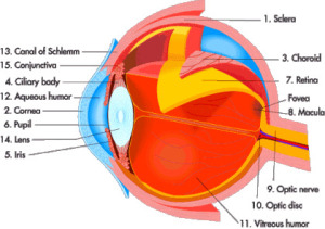
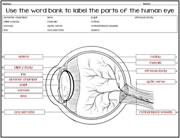
Parts of the eye without labels
Anatomy of the eye: Quizzes and diagrams | Kenhub How to learn the parts of the eye. Found within two cavities in the skull known as the orbits, the eyes are surrounded by several supporting structures including muscles, vessels, and nerves. There are 7 bones of the orbit, two groups of muscles (intrinsic ocular and extraocular), three layers to the eyeball… and that's just the beginning. There's a lot to learn, but stay calm! Shipping Labels, DOT Placards, UN Packaging from ... Find all you need for hazmat shipping. Labelmaster offers UN packaging, CHEMTREC labels, GHS training, CFR's, DG shipping software, hazmat labels and placards and more. Eye Diagram Teaching Resources | Teachers Pay Teachers The Human Eye Overview Reading Comprehension and Diagram Worksheet. by. Teaching to the Middle. 4.7. (65) $1.50. Zip. This passage briefly describes the human eye (900-1000 Lexile). 14 questions (matching and multiple choice) assess students' understanding. Students label a diagram of 6 parts of the eye.
Parts of the eye without labels. Parts of the Microscope with Labeling (also Free Printouts) 5. Knobs (fine and coarse) By adjusting the knob, you can adjust the focus of the microscope. The majority of the microscope models today have the knobs mounted on the same part of the device. Image 5: The circled parts of the microscope are the fine and coarse adjustment knobs. Picture Source: bp.blogspot.com. PDF Eye Anatomy Handout - National Institutes of Health Here are descriptions of some of the main parts of the eye: Cornea: The cornea is the clear outer part of the eye's focusing system located at the front of the eye. Iris: The iris is the colored part of the eye that regulates the amount of light entering the eye. Lens: The lens is a clear part of the eye behind the iris that helps to Eye - Wikipedia Eyes are organs of the visual system.They provide living organisms with vision, the ability to receive and process visual detail, as well as enabling several photo response functions that are independent of vision.Eyes detect light and convert it into electro-chemical impulses in neurons.In higher organisms, the eye is a complex optical system which collects light from the surrounding ... Eye Anatomy: 16 Parts of the Eye & Their Functions - Vision Center The following are parts of the human eyes and their functions: 1. Conjunctiva. The conjunctiva is the membrane covering the sclera (white portion of your eye). The conjunctiva also covers the interior of your eyelids. Conjunctivitis, often known as pink eye, occurs when this thin membrane becomes inflamed or swollen. Other eye disorders that affect the conjunctiva include:
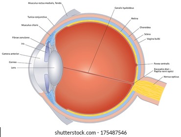
› resources › labelfinderLabelmaster's Hazard Class Label Finder - Hazmat Shipping ... PVC-Free film hazmat labels meet the IMO 90-day sea water immersion test specifications in section 5.2.2.2.1.7. We offer much more than standard DOT hazard class labels. We also provide blank and pre-printed stock proper shipping name labels and international wordless hazmat labels within each hazard class category. Label Resources: en.wikipedia.org › wiki › Human_eyeHuman eye - Wikipedia The human eye is a sensory organ, part of the sensory nervous system, that reacts to visible light and allows us to use visual information for various purposes including seeing things, keeping our balance, and maintaining circadian rhythm.. The eye can be considered as a living optical device.It is approximately spherical in shape, with its outer layers, such as the outermost, white part of ... Anatomy of the Eye | Johns Hopkins Medicine Cornea. The clear, dome-shaped surface that covers the front of the eye. Iris. The colored part of the eye. The iris is partly responsible for regulating the amount of light permitted to enter the eye. Lens (also called crystalline lens). The transparent structure inside the eye that focuses light rays onto the retina. Lower eyelid. Structure and Functions of Human Eye with labelled Diagram - BYJUS The internal components of the eye include: Lens Retina Aqueous humour Optic nerve Vitreous humour Test your knowledge on Structure Of Eye Put your understanding of this concept to test by answering a few MCQs. Click 'Start Quiz' to begin! Select the correct answer and click on the "Finish" button Check your score and answers at the end of the quiz
Parts of the Eye and Their Functions - Robertson Opt Pupil. The pupil appears as a black dot in the middle of the eye. This black area is actually a hole that takes in light so the eye can focus on the objects in front of it. Iris. The iris is the area of the eye that contains the pigment which gives the eye its color. Eye Diagram With Labels and detailed description - BYJUS Cornea is a dome-shaped tissue covering the front of the eye. Iris is the coloured part of the eye and controls the amount of light entering the eye by regulating the size of the pupil. The lens is located just behind the iris. Its function is to focus the light on the retina. The optic nerve transmits electrical signals from the retina to the brain. Anatomy of the eye - Moorfields Eye Hospital Optic nerve: leaves the eye at the optic disc and transfers all the visual information to the brain. Sclera: the white part of the eye, a tough covering with which the cornea forms the external protective coat of the eye. Rod cells are one of the two types of light-sensitive cells in the retina of the eye. There are about 125 million rods ... Eye Pictures, Anatomy & Diagram | Body Maps - Healthline Pads of fat and the surrounding bones of the skull protect them. The eye has several major components: the cornea, pupil, lens, iris, retina, and sclera. These work together to capture an image ...

label the eye quiz Gallery
Eye Anatomy: A Closer Look At the Parts of the Eye - All About Vision For more details about specific structures of the eye and how they function, visit these pages: Conjunctiva Of The Eye. Sclera: The White Of The Eye. Cornea Of The Eye. The Uvea Of The Eye. Pupil: Aperture Of The Eye. The Retina: Where Vision Begins. Macula Lutea Of The Eye. Choroid Of The Eye. Lens Of The Eye. Ciliary Body. Eye Muscles. Aqueous Humor. Optic Nerve
Labeled Parts Of The Eye - ClipArt Best
idlabelinc.com › common-types-warehouse-labelsCommon Types of Warehouse Labels - ID Label Inc. Warehouse Tote and Bin Labels. Warehouses commonly store individual products and parts in plastic bins or containers. Like warehouse racks, these bins should be properly identified with barcode labels to help workers easily locate items, fulfill orders and manage product inventory. Similarly, reusable warehouse totes require proper identification.
5 Best Images of Frog Anatomy Worksheet Answers - Fetal Pig Labeled Diagram, Heart Anatomy ...
What Does the Eye Look Like? - Harvard Eye Associates Cornea: The clear, dome-shaped tissue covering the front of the eye. Fovea: A tiny pit located in the macula of the retina that provides the clearest vision of all. Iris: The colored part of the eye that controls the amount of light that enters the eye by changing the size of the pupil.

Pin on Ylli
The Eyes (Human Anatomy): Diagram, Optic Nerve, Iris, Cornea ... - WebMD The front part (what you see in the mirror) includes: Iris: the colored part; Cornea: a clear dome over the iris; Pupil: the black circular opening in the iris that lets light in

Learn the Nine Essential Parts of Eyeglasses 1. Rims The rims lend form and character to your eyeglasses—they also provide function by holding the lenses in place. 2. End pieces The end pieces are the small parts on the frame that extend outward and connect the lenses to the temples. 3. Bridge The bridge is the center of the frame that rests on your nose and joins the two rims together. 4.

Quiz: Label The Parts Of The Eye - ProProfs Quiz E.Retina A. Optic Nerve B. Iris C. Sclera D. Lens E. Retina

Anatomy of the Eye | Kellogg Eye Center | Michigan Medicine The nerve layer lining the back of the eye. The retina senses light and creates electrical impulses that are sent through the optic nerve to the brain. Sclera The white outer coat of the eye, surrounding the iris. Vitreous Humor The, clear, gelatinous substance filling the central cavity of the eye. How the Eye Works
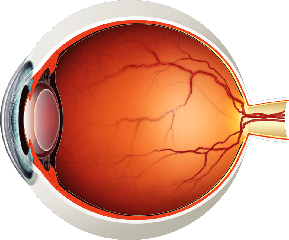
Diagram of the Eye - Lions Eye Institute
Eye in Cross Section : Anatomy : The Eyes Have It - University of Michigan Eye in Cross Section. Click on a label to display the definition. Tap on the image or pinch out and pinch in to resize the image.
Eye Anatomy Labeling Images, Stock Photos & Vectors | Shutterstock
Cornea of the Eye - Definition and Detailed Illustration The cornea is the clear front surface of the eye. It lies directly in front of the iris and pupil, and it allows light to enter the eye. Viewed from the front of the eye, the cornea appears slightly wider than it is tall. This is because the sclera (the "white" of the eye) slightly overlaps the top and bottom of the anterior cornea.
45 Astonishing Facts About Eyes - Information Loaded By HNBT
Your Eyes (for Kids) - Nemours KidsHealth The cornea (say: KOR-nee-uh), a transparent dome, sits in front of the colored part of the eye. The cornea helps the eye focus as light makes its way through. It is a very important part of the eye, but you can hardly see it because it's made of clear tissue. Like clear glass, the cornea gives your eye a clear window to view the world through.

31 Label The Parts Of Eye - Labels For Your Ideas
Parts of the Eye - Chester F. Carlson Center for Imaging Science Iris/Pupil. Iris is heavily pigmented. Sphincter muscle to constrict or dilate the pupil. Pupil is the hole through which light passes. Pupil diameter ranges from about 3-7 mm. Area of 7-38 square mm (factor of 5) Eye color (brown, green, blue, etc.) dependent on amount and distribution of the pigment melanin.

picture front of the eye without labels clipart - Clipground
Eye Anatomy: Parts of the Eye and How We See The surface of the eye and the inner surface of the eyelids are covered with a clear membrane called the conjunctiva. The layers of the tear film keep the front of the eye lubricated. Tears lubricate the eye and are made up of three layers. These three layers together are called the tear film. The mucous layer is made by the conjunctiva.

Medical Stock Art, Anatomy of the Eye
Iris of the Eye: Definition, Anatomy & Common Conditions - Cleveland Clinic The iris is the colored part of your eye. Muscles in your iris control your pupil. ... Some people are born without an iris in one or both of their eyes — a genetic condition called aniridia. Without an iris, your eye would still function, but your vision would be blurry. ... Wear sunglasses with 100% UV protection or a UV400 label anytime ...

Label Parts of the Human Eye - University of Dayton Parts of the Eye. Select the correct label for each part of the eye. The image is taken from above the left eye. Click on the Score button to see how you did. Incorrect answers will be marked in red. ...

Amazon.com: Human Eye Anatomy Medical Chart Educational Diagram Laminated Dry Erase Sign Poster ...
Label the Eye - The Biology Corner Label the Eye. Shannan Muskopf December 30, 2019. This worksheet shows an image of the eye with structures numbered. Students practice labeling the eye or teachers can print this to use as an assessment. There are two versions on the google doc and pdf file, one where the word bank is included and another with no word bank for differentiation.

PDF Parts of the Eye - National Institutes of Health Macula: The macula is the small, sensitive area of the retina that gives central vision. It is located in the center of the retina. Optic nerve: The optic nerve is the largest sensory nerve of the eye. It carries impulses for sight from the retina to the brain. Pupil: The pupil is the opening at the center of the iris.
Label Of Eye - Drawing and labeling of eye outer part #humaneyediagram #fineartsguruji ...
› sailboatSailboat Hardware, Sailboat Accessories & Parts | Fisheries ... Sailboat Hardware, Parts & Accessories Fisheries Supply is your premier supplier of sailboat hardware and accessories from top brands like Harken, Ronstan, Lewmar, Schaefer and more. Both online and in our Seattle store, we offer a full range of quality sailboat supplies including winches, rigging parts, blocks, spinnaker poles, shackles ...
Post a Comment for "38 parts of the eye without labels"