39 human brain with labels
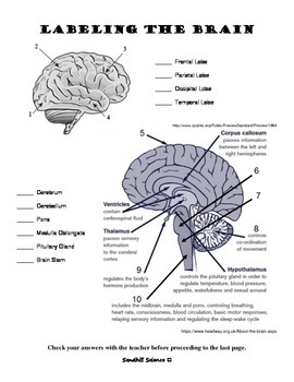
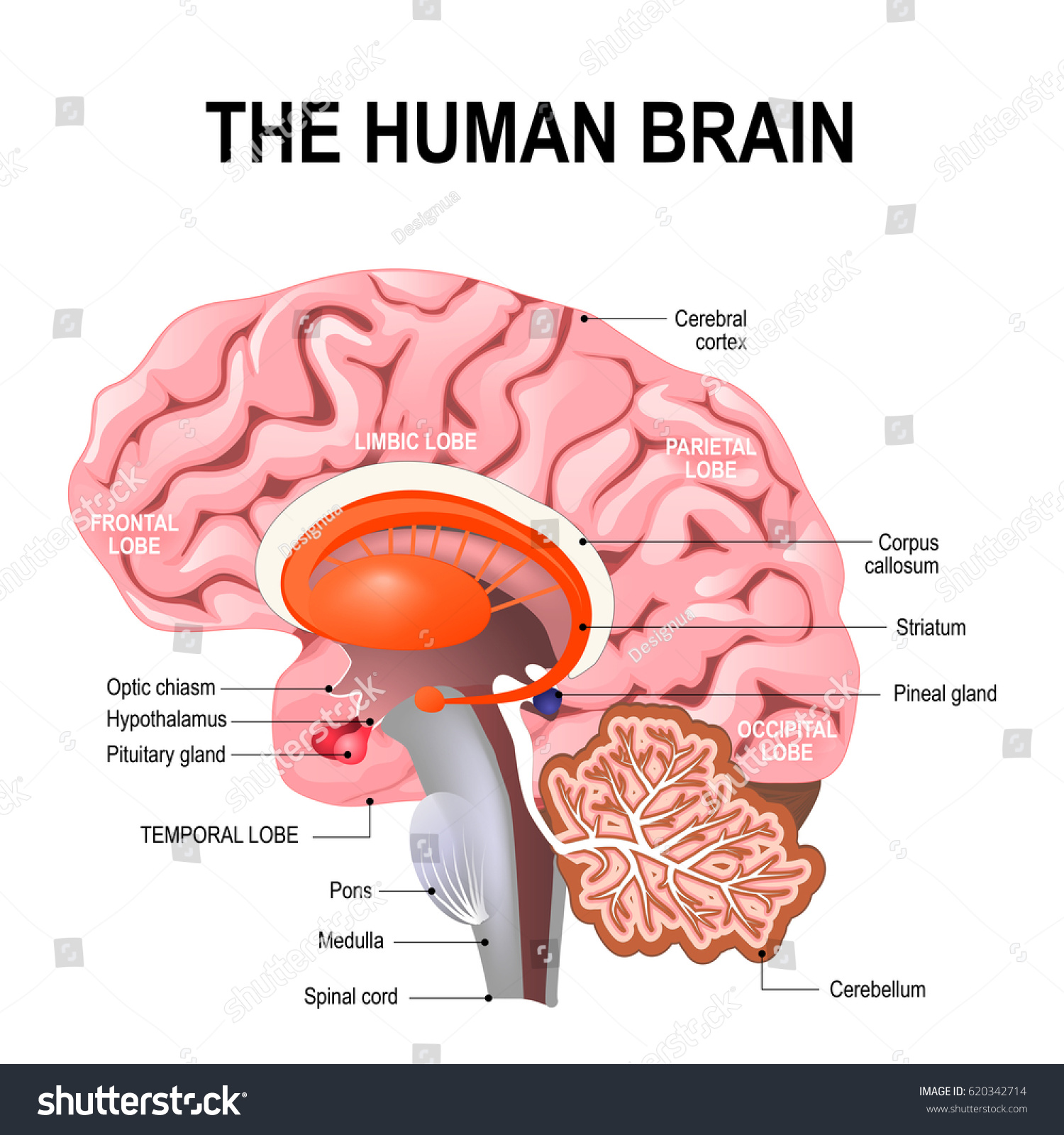
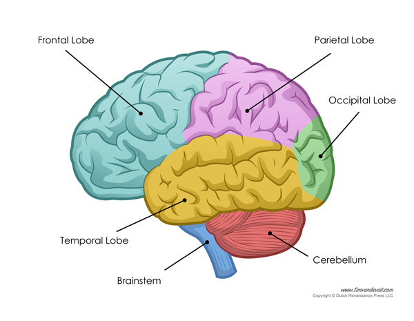
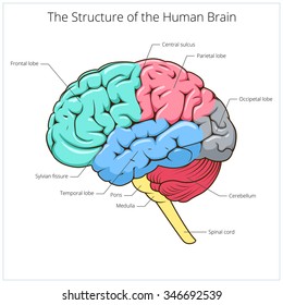
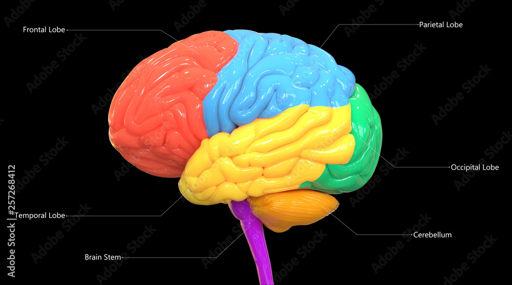
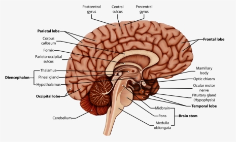
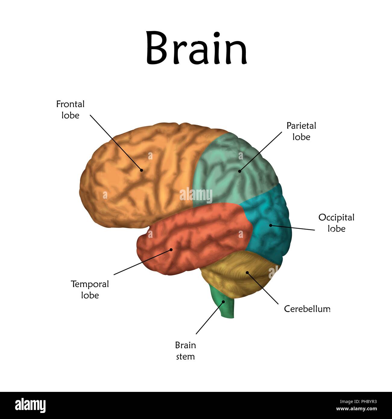
Brain - Human Brain Diagrams and Detailed Information - Innerbody Pons Quadrigeminal Lamina Superior Colliculus Cerebellum Cerebellar Peduncle 4th Ventricle Cerebral Aqueduct Choroid Plexus FOREBRAIN Diencephalon Choroid Plexus of 3rd Ventricle Optic Chiasm Hypothalamic Sulcus Hypothalamus Interthalamic Adhesion Optic Recess Pineal Gland Stria Medullaris of Thalamus Thalamus (3rd Ventricle) Tuber Cinereum Parts of the Brain: Structures, Anatomy and Functions The brain is a 3-pound organ that contains more than 100 billion neurons and many specialized areas. There are 3 main parts of the brain include the cerebrum, cerebellum, and brain stem.The Cerebrum can also be divided into 4 lobes: frontal lobes, parietal lobes, temporal lobes, and occipital lobes.The brain stem consists of three major parts: Midbrain, Pons, and Medulla oblongata.
Human brain with labels , black and white royalty-free images Find Human brain with labels , black and white stock images in HD and millions of other royalty-free stock photos, illustrations and vectors in the Shutterstock collection. Thousands of new, high-quality pictures added every day.

Human brain with labels
The Human Brain Atlas - Michigan State University The Human Brain Atlas Keith D. Sudheimer, Brian M. Winn, Garrett M. Kerndt, Jay M. Shoaps, Kristina K. Davis, Archibald J. Fobbs Jr., and John I. Johnson Radiology Department, Communications Technology Laboratory, and College of Human Medicine, Michigan State University; National Museum of Health and Medicine, Armed Forces Institute of Pathology Main Parts of the Human Brain and Subdivisions of Human Brain Parts Thalamus, epithalamus, subthalamus and hypothalamus are the four sub-divisions. Here Hypothalamus is one of the parts of the human brain that initiates, coordinates, maintains and assists in the successful accomplishment of a number of visceral activities with the help of its hormonal secretions. Moreover, the sensory Optic Nerve, coming from ... human brain with labels MRI Brain Vascular Anatomy | Mri Scan Images | Mri brain, Mri, Thrombosis. 9 Images about MRI Brain Vascular Anatomy | Mri Scan Images | Mri brain, Mri, Thrombosis : Superior view of human brain with colored lobes and labels Stock Photo, Living in a Brainstorm: Gushy Brains! and also Occipital Bone of the Human Skull | ClipArt ETC. MRI Brain ...
Human brain with labels. Amazon.com: XINDAM 3D Human Brain with Labels Anatomical Model ... XINDAM 3D Human Brain with Labels Anatomical Model Paperweight (Laser Etched) in Crystal Glass Ball Science Gift (Included LED Base) Brand: XINDAM 30 ratings $6699 FREE Returns Coupon: Save an extra 5% when you apply this coupon Terms Size:3.2 Inch Made from glass and the amazing power of a laser. Brain (Human Anatomy): Picture, Function, Parts, Conditions ... - WebMD Glioblastoma: An aggressive, malignant brain tumor (cancer). Brain glioblastomas progress rapidly and are very difficult to cure. Hydrocephalus: An abnormally increased amount of cerebrospinal... Labeled Diagrams of the Human Brain You'll Want to Copy Now Labeled Diagrams of the Human Brain Central Core The central core consists of the thalamus, pons, cerebellum, reticular formation and medulla. These five regions are the central areas that regulate breathing, pulse, arousal, balance, sleep and early stages of processing sensory information. Labeled Brain Model Diagram | Science Trends The cerebrum is the largest and most complex portion of the human brain. The cerebrum's function is to control our actions and thoughts, either conscious or unconscious, and responses to stimuli. The cerebrum itself is typically divided into four different lobes: the temporal lobe, the parietal lobe, the occipital lobe, and the frontal lobe.
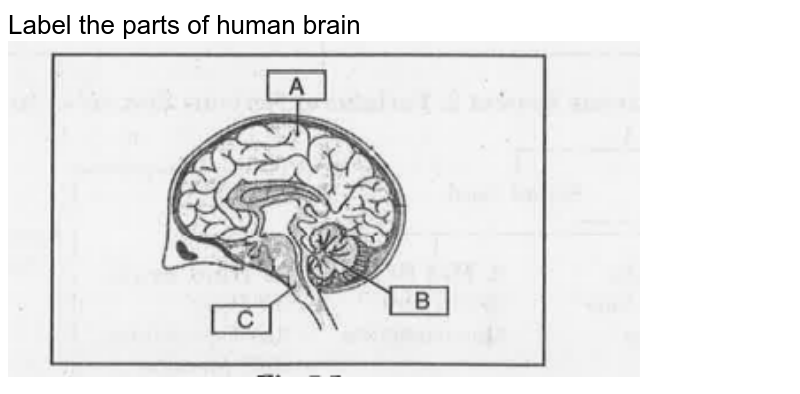
Brain: Atlas of human anatomy with MRI - e-Anatomy - IMAIOS Anatomy of the brain (MRI) - cross-sectional atlas of human anatomy. The module on the anatomy of the brain based on MRI with axial slices was redesigned, having received multiple requests from users for coronal and sagittal slices. The elaboration of this new module, its labeling of more than 524 structures on 379 MRI images in three different ... Human brain - Wikipedia The cerebrum, the largest part of the human brain, consists of two cerebral hemispheres. Each hemisphere has an inner core composed of white matter, and an outer surface - the cerebral cortex - composed of grey matter. The cortex has an outer layer, the neocortex, and an inner allocortex. Human Brain Photos and Premium High Res Pictures - Getty Images Browse 28,900 human brain stock photos and images available, or search for human brain anatomy or human brain illustration to find more great stock photos and pictures. Related searches: human brain anatomy human brain illustration human brain diagram human brain scan human brain vector of 100 NEXT Labeling the Brain | Human Anatomy Quiz - Quizizz answer choices Cerebrum Cerebellum Brain Stem Diencephalon Question 16 120 seconds Q. Which letter on the illustration represents the frontal lobe ? answer choices A B C D Question 17 120 seconds Q. Which letter on the illustration represents the temporal lobe ? answer choices A B C D Question 18 30 seconds
Human Brain - Structure, Diagram, Parts Of Human Brain - BYJUS Following are the major parts of the human brain: Forebrain - Largest part of the brain It is the anterior part of the brain. The forebrain parts include: Cerebrum Hypothalamus Thalamus Forebrain Function: Controls the reproductive functions, body temperature, emotions, hunger and sleep. Fact: The largest among the forebrain parts is the cerebrum. 109 Labeled Human Brain Illustrations & Clip Art - iStock detailed anatomy of the human brain. Illustration showing the medulla, pons, cerebellum, hypothalamus, thalamus, midbrain. Sagittal view of the brain. Isolated on a white background. Cerebrospinal fluid vector illustration. Anatomical labeled... Cerebrospinal fluid vector illustration. Anatomical labeled scheme with human head and inside of skull. Nervous System - Label the Brain - TheInspiredInstructor.com Nervous System - Label the Brain Nervous System - Brain Name: Choose the correct names for the parts of the brain. ( 1) (2) (3) (4) (5) (6) (7) (8) ( 9) This brain part controls thinking. (10) This brain part controls balance, movement, and coordination. (11) This brain part controls involuntary actions such as breathing, heartbeats, and digestion. Human Brain Anatomy - Components of Human Brain with Images i. Cerebrum: The Largest Part Composed of the right and left hemispheres, it is the largest part of the brain and is responsible for the processing of speech, learning, reasoning, emotions, muscular contractions as well as the interpretation of sensory data related to hearing, vision and touch. ii. Cerebellum—the Sub-Cerebral Region:
Amazon.com: Labeled Brain Model Axis Scientific Human Brain Model Anatomy with Colored and Labeled Regions, 2-Part Human Brain Model Disassembled - Includes Base, Detailed Product Manual and 3 Year Warranty 16 $18799 FREE delivery Tue, Oct 25 Or fastest delivery Mon, Oct 24 Small Business
Human Brain Model Making Using Cardboard With Labels | DIY ... Human Brain Model Making Using Cardboard With Labels | DIY | craftpiller#humanbrainmodel #usingcardboard #craftpiller #diy #biologymodel #zoologymodel #scien...
Human Brain Labels Pictures, Images and Stock Photos Triune brain: Reptilian complex (basal ganglia for instinctual behaviours), mammalian brain (septum, amygdalae, hypothalamus, hippocamp for feeling) and Neocortex (cognition, language, sensory perception, and spatial reasoning). Cross section of the human brain. Vector illustration for medical, biological, educational and science use
Stock Images, Photos, Vectors, Video, and Music | Shutterstock Stock Images, Photos, Vectors, Video, and Music | Shutterstock
Brain Anatomy and How the Brain Works | Johns Hopkins Medicine The occipital lobe is the back part of the brain that is involved with vision. Temporal lobe. The sides of the brain, temporal lobes are involved in short-term memory, speech, musical rhythm and some degree of smell recognition. Deeper Structures Within the Brain Pituitary Gland
Human Brain Diagram - Labeled, Unlabled, and Blank - Pinterest Learn the parts of the human brain with these convenient printables for students and teachers. Pinterest. Today. Watch. Explore. When autocomplete results are available use up and down arrows to review and enter to select. Touch device users, explore by touch or with swipe gestures. ... Human Brain Diagram - Labeled, Unlabled, and Blank ...
Parts of the brain: Learn with diagrams and quizzes | Kenhub Labeled brain diagram. First up, have a look at the labeled brain structures on the image below. Try to memorize the name and location of each structure, then proceed to test yourself with the blank brain diagram provided below. Labeled diagram showing the main parts of the brain.
Diagram Of Brain with their Labelings and Detailed Explanation - BYJUS The human brain is divided into three main parts: Forebrain. Midbrain. Hindbrain. These three main parts comprises many small parts. Forebrain The forebrain is also called as Prosencephalon. The forebrain is the anterior part of the brain, which comprises the cerebral hemispheres, the thalamus, and the hypothalamus.

BEAMNOVA Human Brain Model 2 Times Life Size for Neuroscience Teaching with Labels Anatomy Model for Learning Science Classroom Study Display Medical ...
The Human Brain - Visible Body Rotate this 3D model to see the four major regions of the brain: the cerebrum, diencephalon, cerebellum, and brainstem. The brain directs our body's internal functions. It also integrates sensory impulses and information to form perceptions, thoughts, and memories. The brain gives us self-awareness and the ability to speak and move in the world.
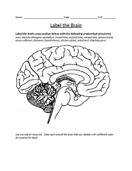
brain label worksheet Label The Human Brain View - 4th Grade Science Worksheet - SoD . sod schoolofdragons. Nervous System Worksheet - WikiEducator . diagram neuron unlabeled worksheet system nerve cell nervous function kinds different muscle three wikieducator impulse anatomy neurone fibres match functions.
3D Brain 3D Brain This interactive brain model is powered by the Wellcome Trust and developed by Matt Wimsatt and Jack Simpson; reviewed by John Morrison, Patrick Hof, and Edward Lein. Structure descriptions were written by Levi Gadye and Alexis Wnuk and Jane Roskams. Copyright © Society for Neuroscience (2017).
human brain with labels MRI Brain Vascular Anatomy | Mri Scan Images | Mri brain, Mri, Thrombosis. 9 Images about MRI Brain Vascular Anatomy | Mri Scan Images | Mri brain, Mri, Thrombosis : Superior view of human brain with colored lobes and labels Stock Photo, Living in a Brainstorm: Gushy Brains! and also Occipital Bone of the Human Skull | ClipArt ETC. MRI Brain ...
Main Parts of the Human Brain and Subdivisions of Human Brain Parts Thalamus, epithalamus, subthalamus and hypothalamus are the four sub-divisions. Here Hypothalamus is one of the parts of the human brain that initiates, coordinates, maintains and assists in the successful accomplishment of a number of visceral activities with the help of its hormonal secretions. Moreover, the sensory Optic Nerve, coming from ...
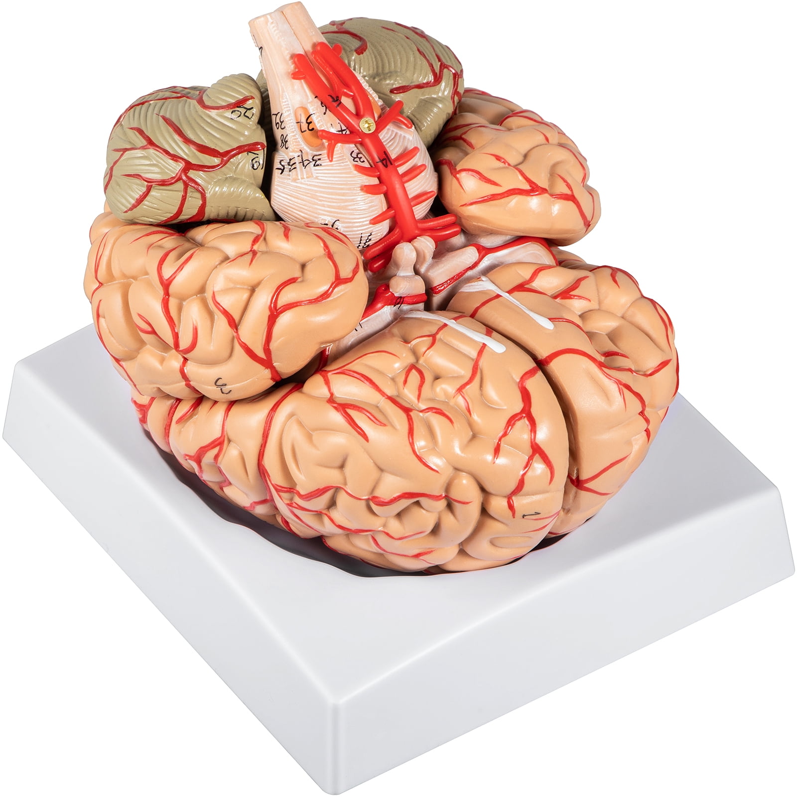
VEVOR Human Brain Model Anatomy 9-Part Model of Brain W/Labels & Display Base Color-Coded Life Size Human Brain Anatomical Model Brain Teaching Tool ...
The Human Brain Atlas - Michigan State University The Human Brain Atlas Keith D. Sudheimer, Brian M. Winn, Garrett M. Kerndt, Jay M. Shoaps, Kristina K. Davis, Archibald J. Fobbs Jr., and John I. Johnson Radiology Department, Communications Technology Laboratory, and College of Human Medicine, Michigan State University; National Museum of Health and Medicine, Armed Forces Institute of Pathology

XINDAM 3D Human Brain with Labels Anatomical Model Paperweight(Laser Etched) in Crystal Glass Ball Science Gift (Included LED Base)


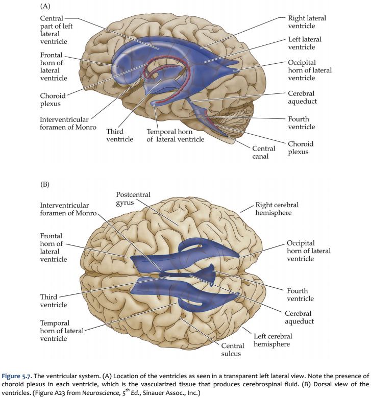


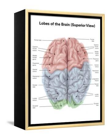
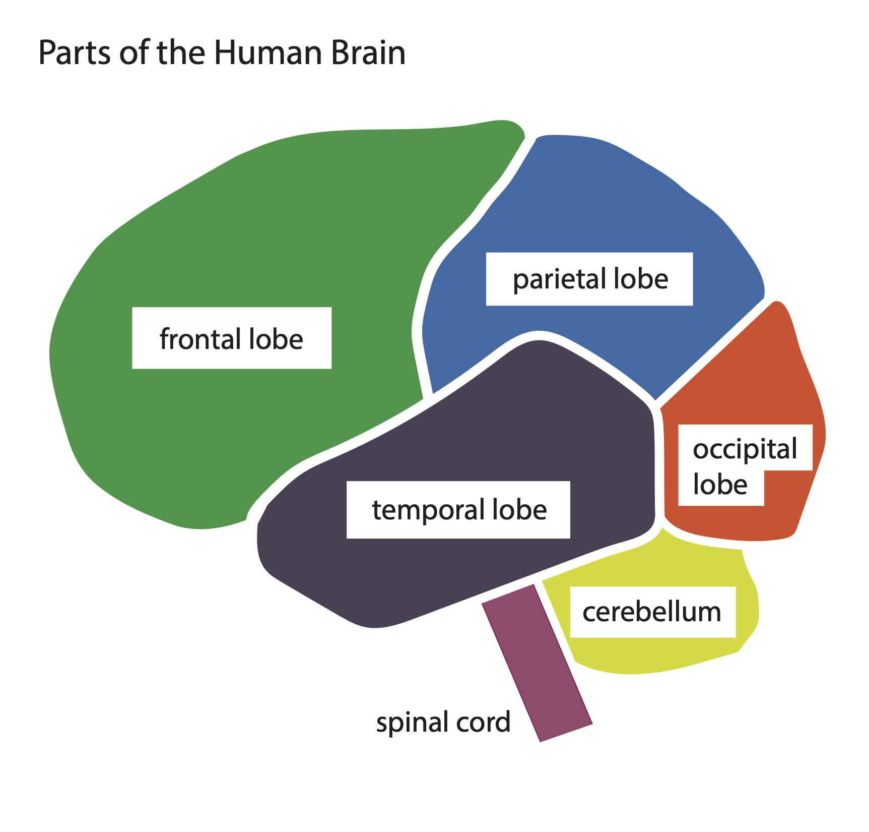










Post a Comment for "39 human brain with labels"